Splice Site Boundaries
Extension of reading frame for length conservation
Found on contig47 of the D. mojavensis dot, rho-5 is a weakly conserved gene compared to its D. melanogaster ortholog. We will try to annotate the penultimate exon (CDS 5_9866_0) in this example.
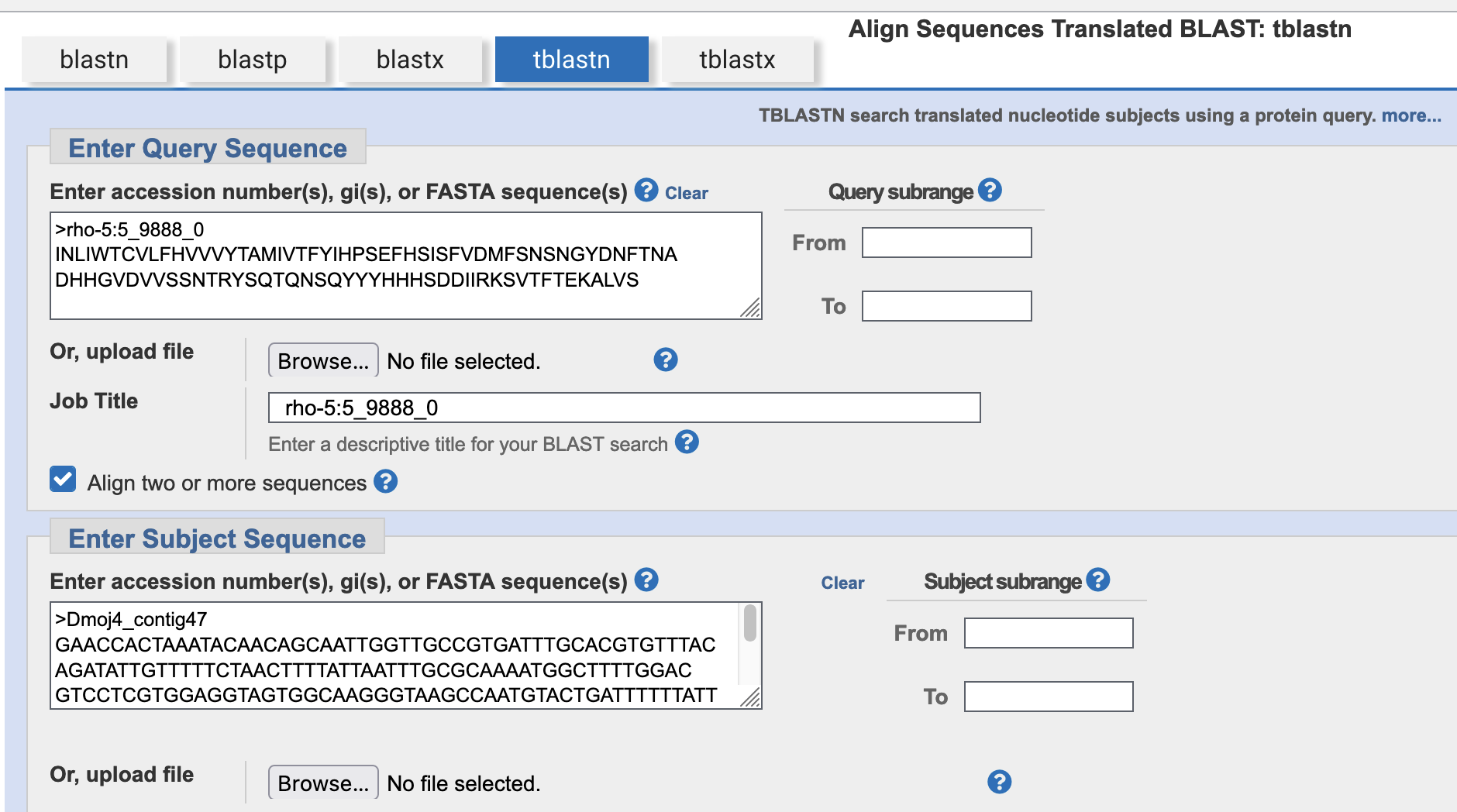
Step 1: Using the amino acid sequence from this exon as the query and the nucleotide sequence of contig47 as the subject, perform a tblastn search with the low complexity filter and compositional adjustment turned off (Figure 1).
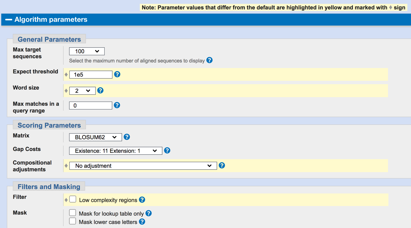
If you cannot find any alignments using these search parameters, you can increase the sensitivity of the BLAST search by changing the following parameters (Figure 2):
-
Increase the Expect value threshold from 0.05 to 1e5 or more.
-
Decrease the word size from 5 to 3 (database searches) or 3 to 2 (bl2seq searches).
-
Define a subrange to find the best alignment in a smaller region.
For additional information on word size, compositional adjustment, etc., click on the question mark next to the drop-down box corresponding to the parameter of interests.
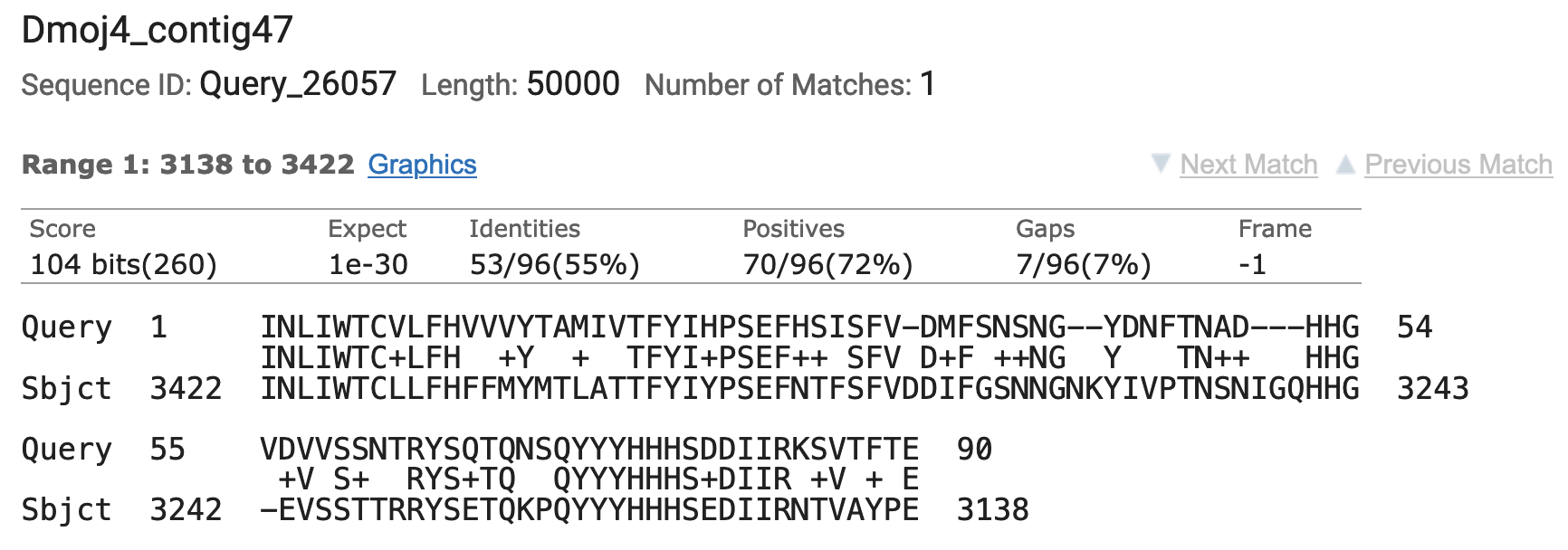
I was able to find a good alignment for the penultimate exon, and it is found within the expected region of contig47 (Figure 3). While the percent identity is only 55%, the alignment accounts for the first 90 residues of this CDS. However, CDS 5_9866_0 is 95 residues long so we are still missing five amino acids at the 3' end.
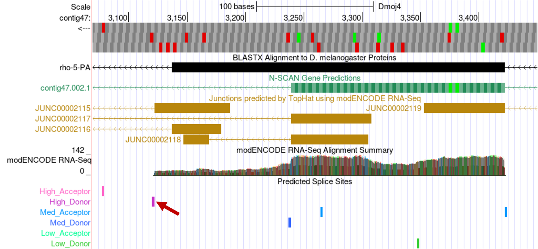
Step 2: Open a web browser and navigate to contig47 on the Genome Browser (D. mojavensis Sep. 2008 assembly) at gander.wustl.edu so that we can take a closer look at this region. Figure 4 shows that there are multiple evidence tracks that support the boundaries for this exon. Data from the modENCODE RNA-Seq coverage, TopHat junctions, and donor/acceptor splice site tracks all support this region very well.
While there are several TopHat junctions that suggest the exon should be shorter than the boundaries suggested by the RNA-Seq read coverage data, using these splice sites would remove some of the conserved amino acids of CDS 5_9866_0. For example, if we were to follow the N-SCAN prediction, much of the conserved sequence would be truncated. This model would also contradict the high-scoring donor site predicted right where the RNA-Seq data stops.
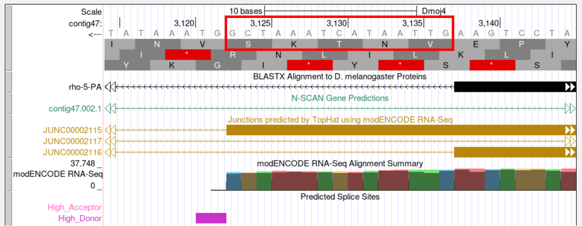
Zooming in on the 3' end, we found the donor site at 3121-3122. This will extend the aligned region and gives us the five amino acids that were missing from the tblastn alignment (Figure 5).
Therefore, the final exon coordinates for CDS 5_9866_0 should be 3422-3123 in order to preserve the conserved amino acids and to minimize the number of changes compared to the D. melanogaster gene model.
Selecting a better supported splice site
While extension of an open reading frame is important for conserving the length of the coding exon, it is also important to consider selecting the best-supported splice site. For example, let’s look at the first coding exon (1_2333_0) of Dyrk3 in D. mojavensis.
Found on contig37, the blastx track on the Genome Browser reveals an alignment that only covers a small part of the first exon. Consequently, we will need to do a more sensitive tblastn search to determine if there is any additional region of conservation to the first exon. The second exon of Dyrk3 begins at 55,082. Because this gene is oriented in the positive direction, we will look for an exon upstream from this coordinate.
Step 1: Perform a tblastn search using the amino acid sequence for the first coding exon of Dyrk3 (CDS 1_2333_0) as the query and the contig37 nucleotide sequence as the subject with the low complexity filter and compositional adjustments turned off and the Expect threshold set to 10. This tblastn search reported a spurious match to the end of the CDS at 14520-14543 (E-value = 7.2).
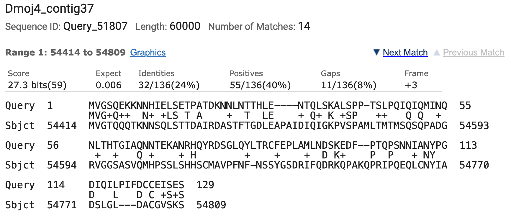
To increase the sensitivity of the tblastn search, I repeated the search using the same parameters except for changing the word size from 3 to 2. This tblastn search with the smaller word size was able to locate an alignment that almost covers the entire first exon (Figure 6). The first exon is 131 aa long and this alignment covers the first 129 aa of this exon.
Step 2: Examining this region in the Genome Browser, we find that this region is located upstream of the second exon and it is well-supported by the RNA-Seq data (Figure 7):
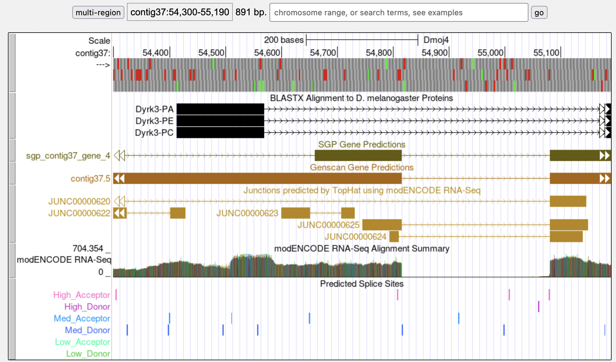
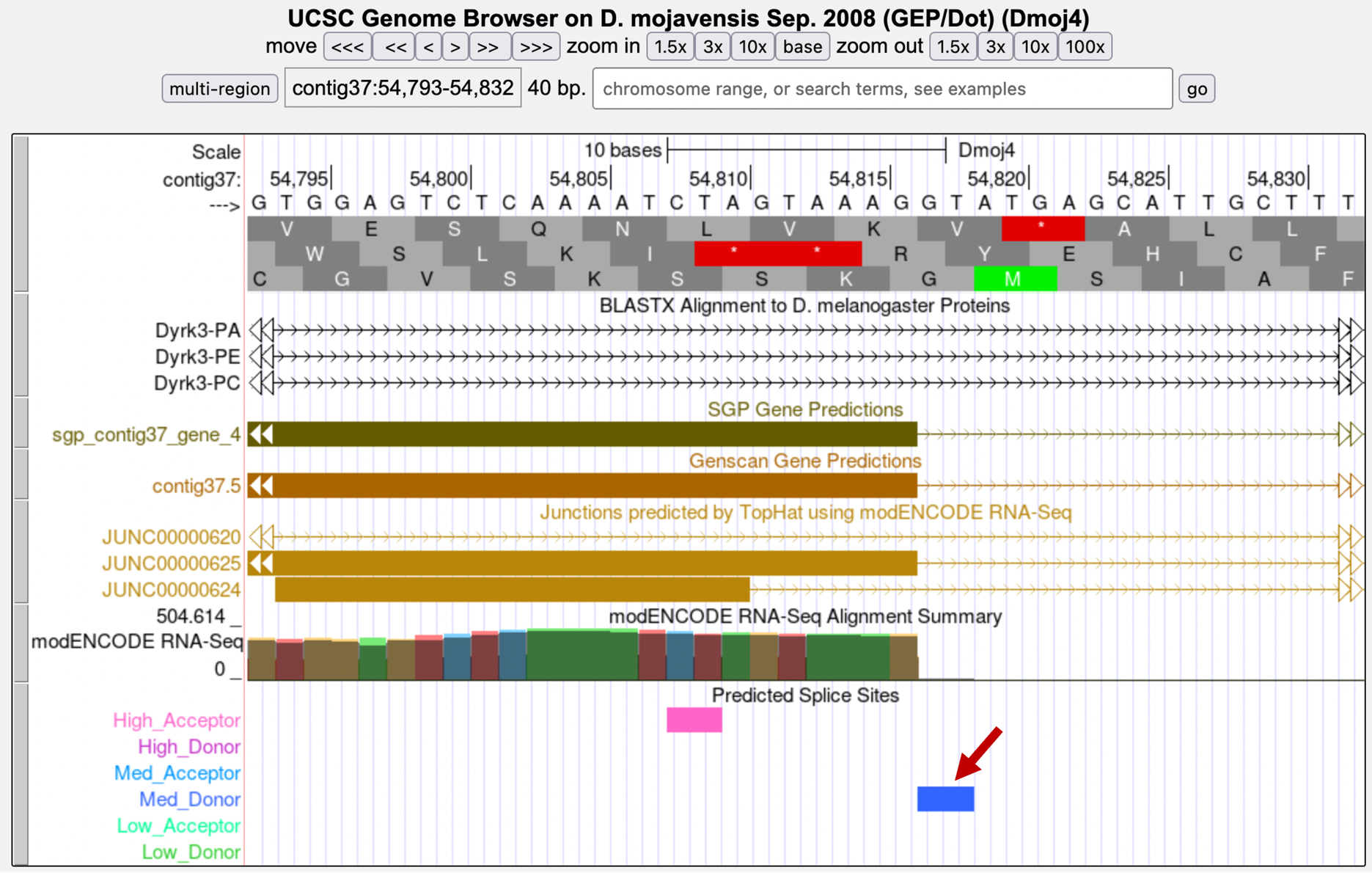
Zooming in on this region, we can see there is some support for 54,810 (where the tblastn alignment terminates) as the end coordinate for the first exon (Figure 8). However, looking a little further downstream, you can see there is much stronger support to place the end of the exon at 54,816. While there are TopHat junctions suggesting both of these splice donor sites are possible (and both junctions have the same splice acceptor site), the splice site at 54,817-54,818 is supported by the SGP and Genscan gene predictions, the modENCODE RNA-Seq data, and a medium quality splice donor site predicted by GeneSplicer.
Collectively, the available evidence provides better support for the donor site at 54,817-54,818 than the donor site located near the end of the tblastn alignment at 54,811-54,812. Selecting this site will also account for the two amino acids length difference between the tblastn alignment and the D. melanogaster CDS. Therefore, we will annotate the first exon for Dyrk3 so that it spans from 54,414 to 54,816 (Figure 8).
Missing/Extra Exons
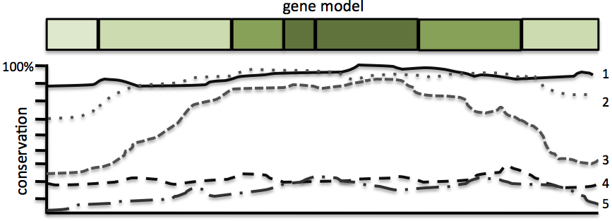
The first and last exons of a gene model are often less conserved. Depending on the conservation of the internal exons, there might be some conservation at the 3' end of the first exon and at the 5' end of the last exon (Figure 9). Sometimes, there will be very high conservation throughout the entire gene model, but this will not always be the case.
Exons found on adjacent contigs
This example will demonstrate the step-by-step strategy for annotating the first coding exon (1_3477_0) of Or13a on contig47 in D. mojavensis. According to the blastx track on the GEP UCSC Genome Browser, Or13a is oriented on the minus strand in contig47. The CDS 1_3477_0 has a total length of 117 amino acids in D. melanogaster.
Step 1: I performed a tblastn search with the CDS 1_3477_0 from Or13a as the query against the nucleotide sequence from contig47 with the low complexity filter and compositional adjustments turned off and the default Expect threshold of 0.05. This search did not produce any significant hits in the correct orientation (i.e., on the minus strand) that contains a large open reading frame with a start codon. Even after modifying the BLAST parameters (i.e., decrease the word size, and increase the E-value threshold) and restricting the subject range to where the first exon should be located, there were only partial low quality matches.
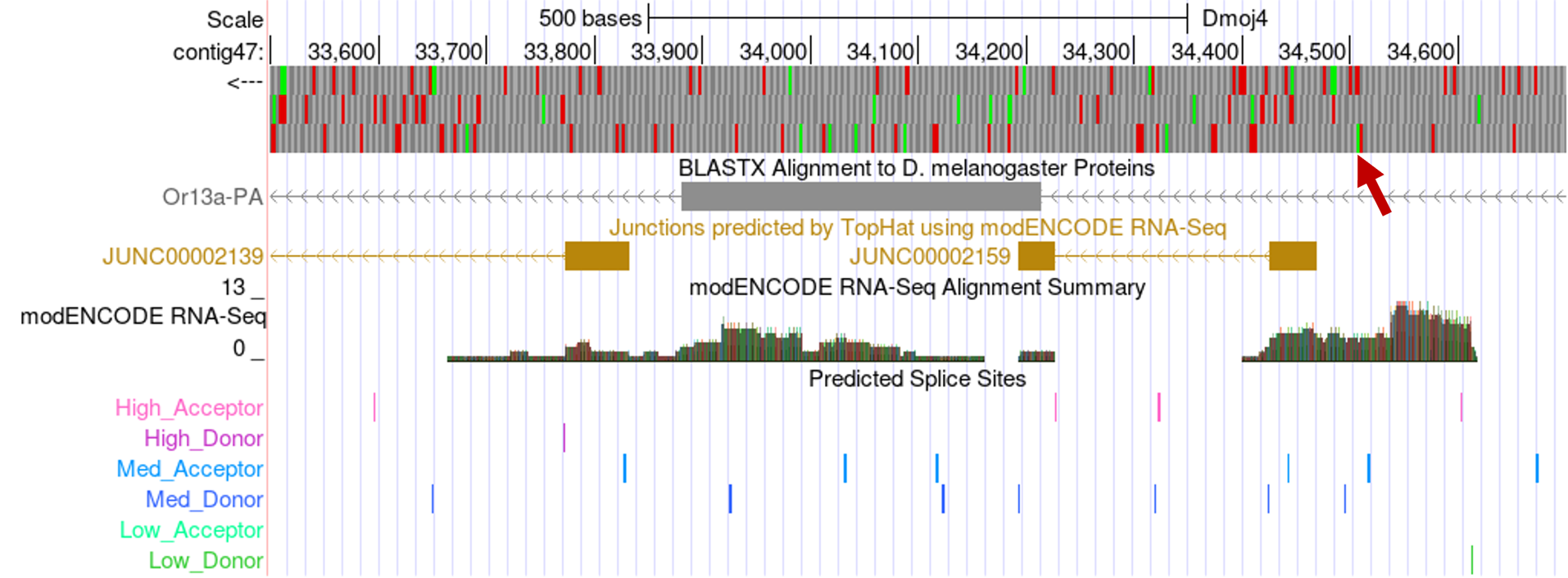
Step 2: Because we cannot place the CDS 1_3477_0 based on the tblastn search results, we will need to rely on the RNA-Seq data to annotate this CDS. Looking at the Genome Browser for contig47, the RNA-Seq data and TopHat junctions suggest that the orthologous CDS might not exist in D. mojavensis.
The TopHat junction JUNC00002159 suggests an intron that spans from 34,425 to 34,227. There is an open reading frame and a start codon (at 34,509-34,507) upstream of 34,425 in frame -3 (Figure 10). However, the splice donor site at 34,424-34,425 is in phase 0 relative to frame -3 and this splice donor site is incompatible with the phase 1 acceptor site at 34,228-34,227 relative to frame -2. Consequently, the open reading frame at 34,509-34,426 cannot be the first CDS of Or13a in D. mojavensis. In order to use this TopHat splice junction, we would need to propose a change in the gene structure so that the second CDS of Or13a (2_3477_1) in D. melanogaster becomes the initial CDS of the Or13a ortholog in D. mojavensis and the regions upstream of the start codon would correspond to the 5' UTR.
While this is a possibility, we would like to create a gene model that minimizes the number of changes (i.e., construct the most parsimonious model) between D. melanogaster and D. mojavensis. Consequently, we will continue to search for a region within contig47 that could be the first exon of Or13a.
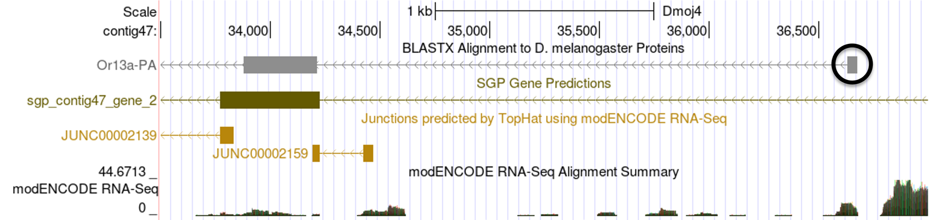
Step 3: We will examine the other evidence tracks on the Genome Browser more closely to see if there are any clues as to the location of the first coding exon. Looking at the region upstream of the start of the second exon, we find a small blastx alignment block for Or13a between 36,500-37,000 (Figure 11):
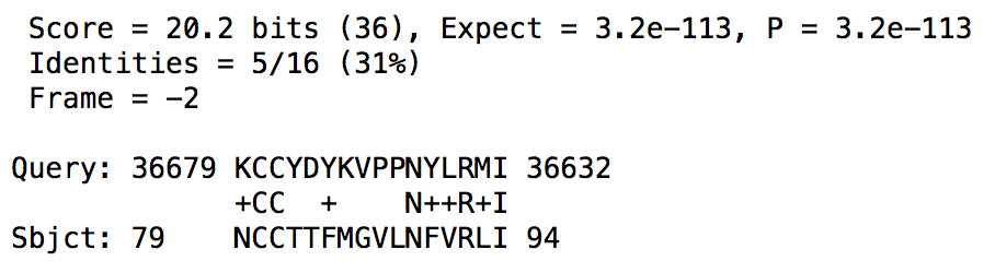
Step 4: Clicking on this blastx block reveals the alignment details for this feature. Organizing the HSP summaries by subject start in ascending order shows that this alignment block spans from 36,679-36,632 in the -2 frame, and it corresponds to residues 79-94 of the CDS (Figure 12). Recall that the first exon of Or13a is 117aa long. This alignment suggests that there is conservation near the end of the first exon with D. melanogaster.
Step 5: Before continuing to examine this region on contig47, it is possible that the first exon is not located within this sequence but could be found on the adjacent contig. A BLAT search of the last 10kb of contig47 against the collection of D. mojavensis dot projects reveals that there is a 20kb overlap between contig47 and contig48. Hence, we will search for the CDS in contig48. If this search did not yield any results, then I would construct the model for the first CDS by extending the open reading frame identified in step 4 to the furthest start codon possible.
However, performing a tblastn search of the first CDS against contig48 with only the low complexity filter and compositional adjustments turned off revealed the following alignment (Figure 13):
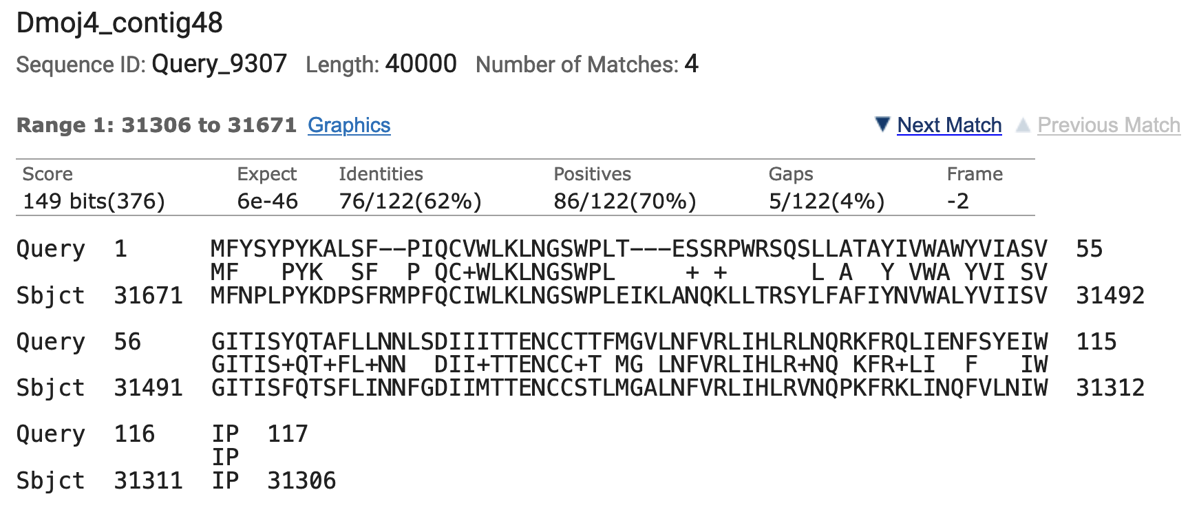
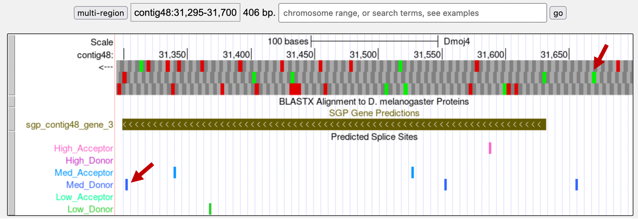
The percent identity for this alignment and the length of sequence conservation is better than all the matches we have previously found on contig47. In addition, there is a medium quality splice donor site at the end of this alignment and an SGP gene prediction (Figure 14). Therefore, we will annotate this region as the first exon of Or13a and make a note in the GEP Annotation Report that the model for this gene on contig47 is a partial annotation that is missing its first exon.
Exons with no sequence conservation
Small exons can be quite difficult to annotate. Below is a step-by-step strategy for annotating the first exon of CG31999 on contig50 in D. mojavensis. CG31999 in D. mojavensis spans five contigs on the forward strand with most of the exons demonstrating moderately strong conservation to the D. melanogaster ortholog.
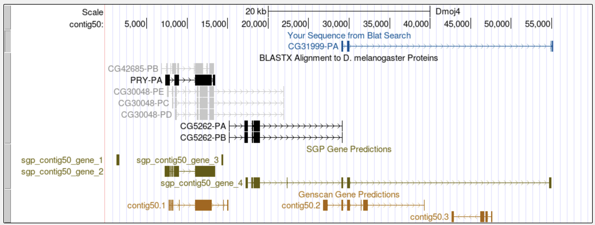
Step 1: We will begin our analysis by performing a BLAT search of the D. melanogaster CG31999-PA protein against the D. mojavensis projects from the dot chromosome (Sep. 2008 assembly). The BLAT result shows that contig50 only contains the first half of CG31999 and CG5262 is the gene that is located closest to the start of CG31999 (Figure 15). A tblastn search of the last CDS of CG5262 (4_7497_2) against contig50 placed this CDS at 18,230-18,985. Examination of the end of the tblastn alignment using the Genome Browser placed the end of the coding region of CG5262 at 18,991.
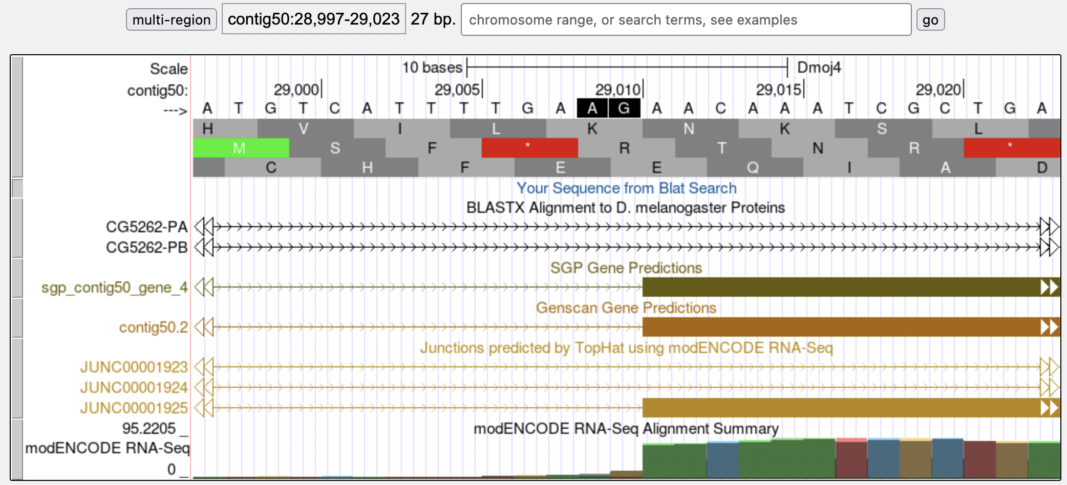
A tblastn search mapped the second CDS of CG31999 (2_10703_2) to 29,016-29,591 in frame +3. (Note that the first amino acid is missing from the alignment, and there is a novel intron at 29,284-29,349.) Examination of this region in the Genome Browser shows the CDS begins at 29,011 and the acceptor site is in phase 2 relative to frame +3 (Figure 16).
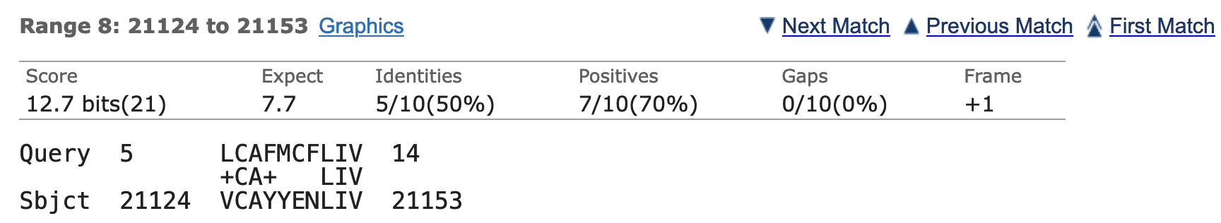
The analysis above allows us to narrow down the location of the first CDS (1_10703_0) to the region between 18,992-29,010. Performing a tblastn search within this subject subrange with the low complexity filter and compositional adjustments turned off and an Expect threshold of 1e5 results in 10 hits to the CDS 1_10703_0 sequence. However, 8 out of the 10 hits are found on the minus strand. The best match (with an E-value of 7.7) on the positive strand is located at 21,124-21,153 in frame +1 (Figure 17).
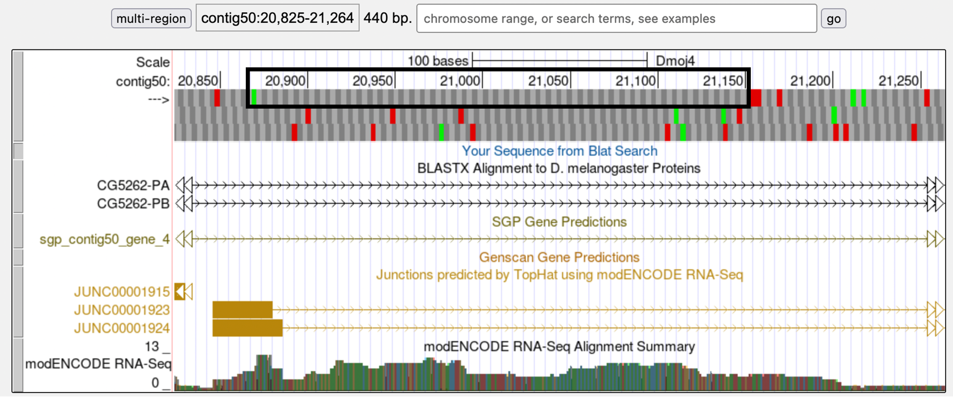
Examination of this region in the Genome Browser shows an open reading frame in frame +1 that is substantially larger (Figure 18) than the expected size of the CDS 1_10703_0 (i.e., 24 amino acids). Consequently, while this region might contain a novel CDS in the D. mojavensis ortholog of CG31999, it is likely not the orthologous CDS of 1_10703_0.
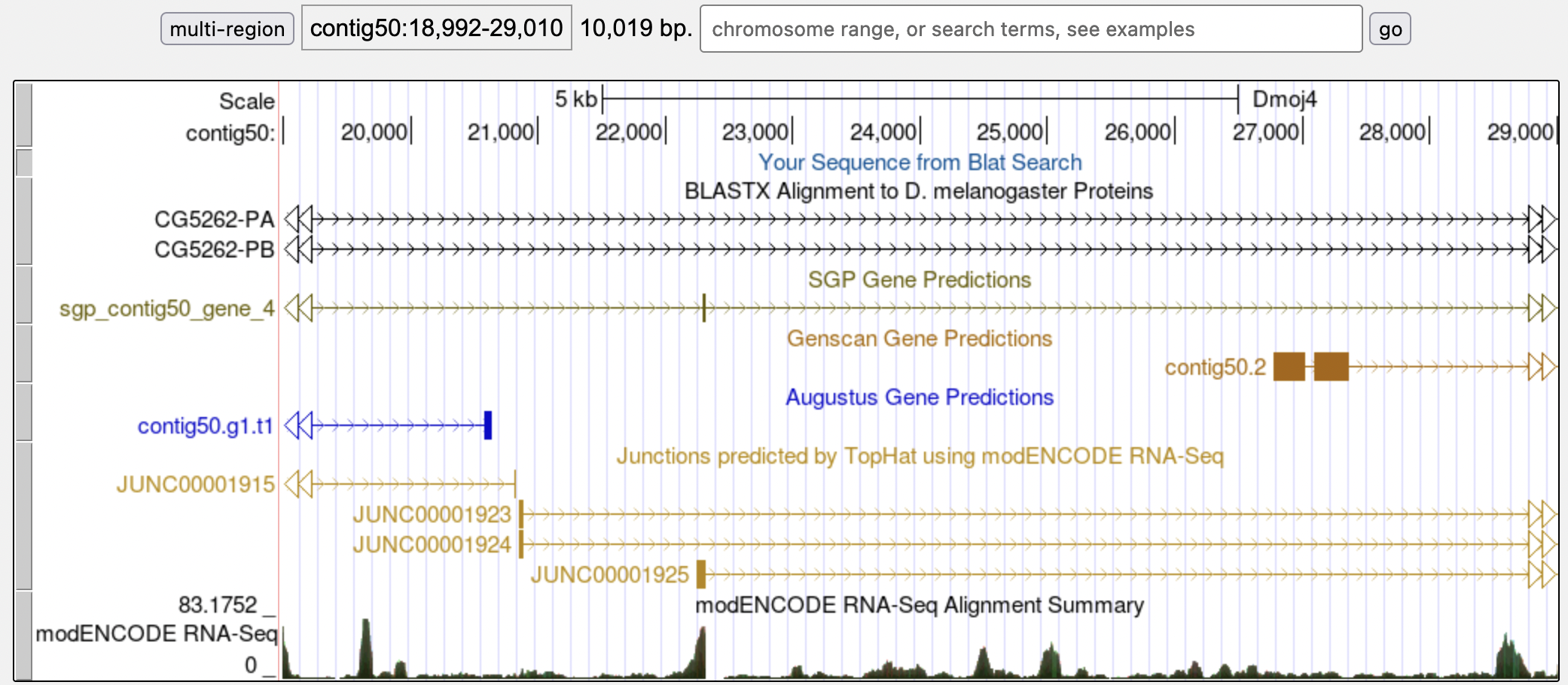
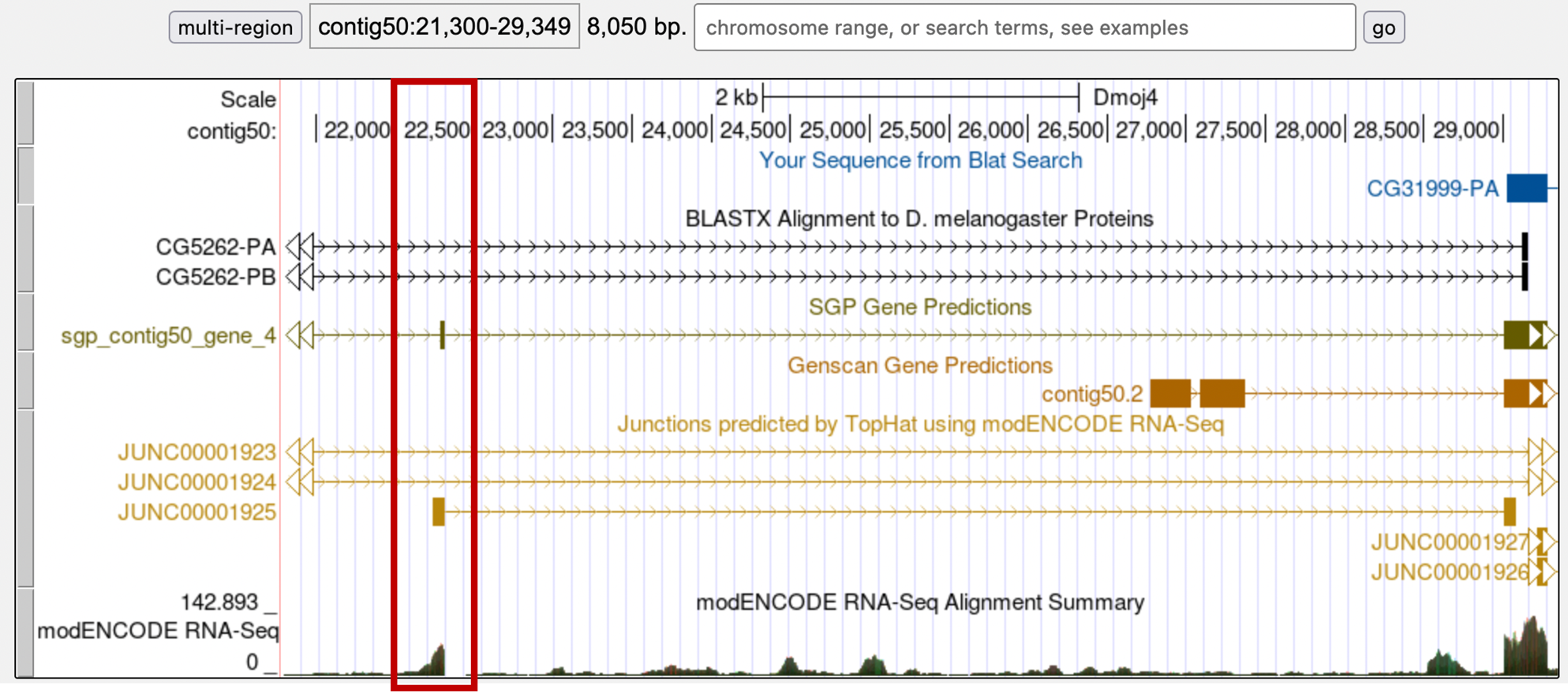
Step 2: Since there is no sequence conservation, perhaps there is some evidence of the first exon in the Genome Browser view of the region. However, unlike contig47, there are very few indicators as to where we could find the CDS 1_10703_0 (Figure 19). While there are some predicted exons from gene predictors, RNA-Seq read coverage, and splice donor site predictions in this region, there is not overwhelming evidence that would favor one specific candidate over other candidates as the putative location of the first exon.
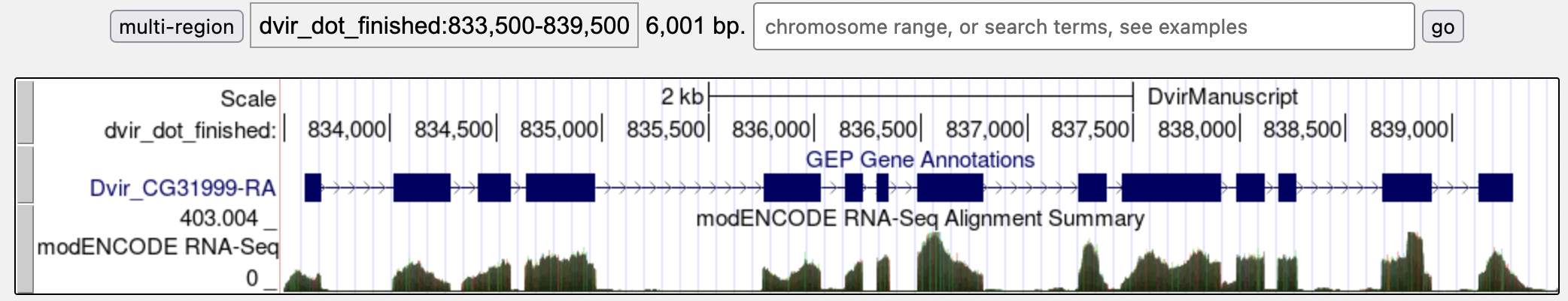
Step 3: With little evidence available in D. mojavensis, D. virilis might provide some additional information. While there is no conservation to the D. melanogaster CDS 1_10703_0, it might be possible to find some conservation with the annotated D. virilis CG31999 model. Going back to the GEP UCSC Genome Browser, I navigated to the region dvir_dot_finished:833,500-839,500 in the D. virilis Manuscript (GEP/2010) assembly (Figure 20). I then click on the CG31999 feature in the GEP Gene Annotations track and then click on the “Translated Protein” link to retrieve the D. virilis CG31999 protein sequence.
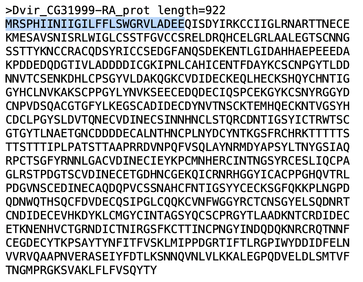
Use the translated D. virilis protein sequence in a blastp alignment against the D. melanogaster amino acid sequence to determine the sequence of the first exon in D. virilis. Figure 21 highlights the region that corresponds to this sequence, which we will use to compare to the D. mojavensis assembly. Unfortunately, this search did not produce any highly significant alignment either.
Step 4: Because there is no sequence conservation at the amino acid level, we will try to find conservation at the nucleotide level through a MUSCLE multiple sequence alignment analysis with D. mojavensis, D. virilis, and D. grimshawi. Any conserved blocks of nucleotide sequence could indicate a potential first exon region.
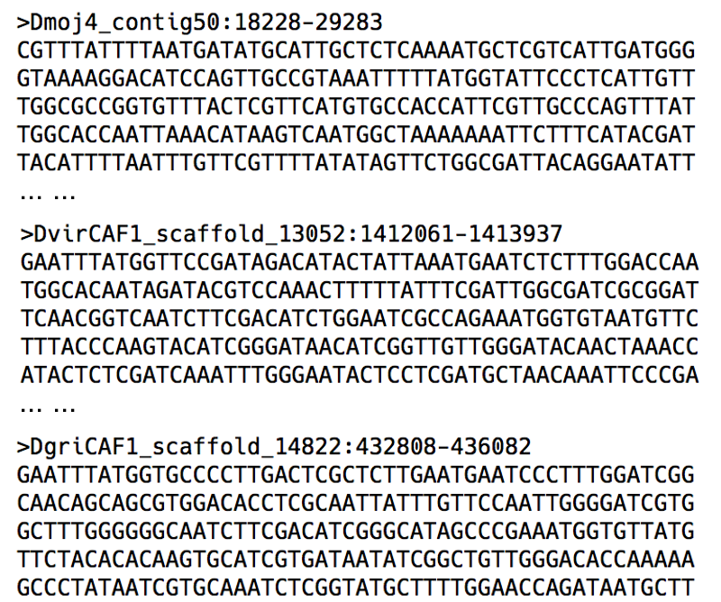
I first extracted the nucleotide sequence from D. mojavensis containing some of the CG5262 and CG31999 flanking sequence in order to anchor the ends of the multiple sequence alignment. I next extracted some of the CG31999 flanking sequence from the D. virilis Manuscript assembly. However, because the 5' end of the orthologous region is flanked by yellow-h instead of CG5262, I cannot use CG5262 to anchor the multiple sequence alignment. (This lack of synteny is also observed in D. grimshawi.)
When I attempt to search for yellow-h on the D. mojavensis dot, there are no hits to this D. melanogaster gene. A FlyBase tblastn search against the D. mojavensis assembly revealed that the putative ortholog of yellow-h in D. mojavensis is found in scaffold_6308. Most of the genes on this D. mojavensis scaffold are orthologous to genes that are found on the D. melanogaster Muller A element (X chromosome).
All of the extracted sequences are saved in a FASTA file (Figure 22) and analyzed by the MUSCLE program. Unfortunately, the MUSCLE alignment revealed no strongly conserved blocks of sequences among the three species.
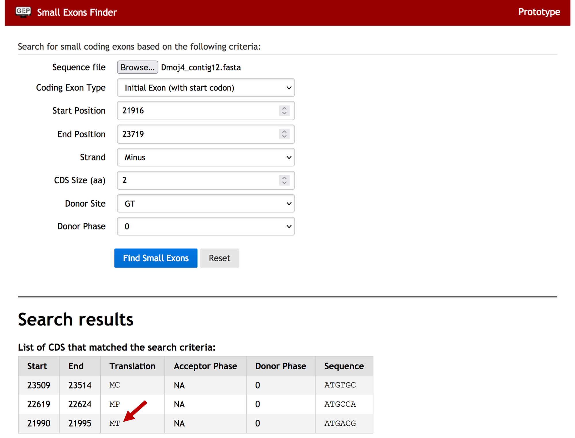
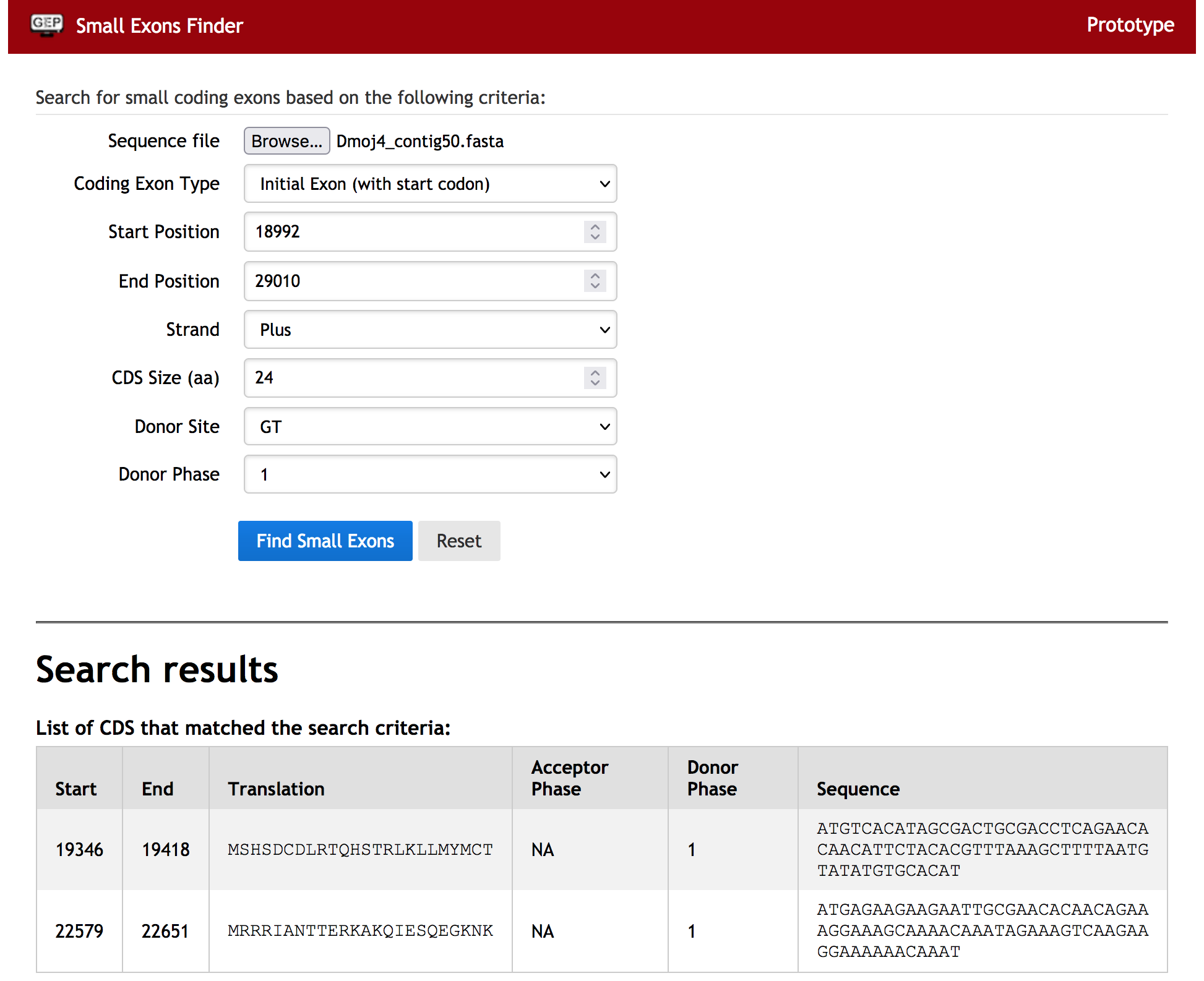
Step 5: With no conservation at any level, we will now try using the Small Exons Finder program to search the contig50 sequence for open reading frames that match the length of the first exon of CG31999. The Small Exons Finder is available through the “Resources & Tools” section of the F Element project page on the GEP website.
Based on our previous analyses, we know that the acceptor site for the second exon is in phase 2, so the donor site for the first exon must be in phase 1. The Small Exons Finder identified two regions that match the search criteria: 19,346-19,418 and 22,579-22,651 (Figure 23). However, these two regions are only weakly supported by the RNA-Seq data. Therefore, I am not inclined to use either of these open reading frames as part of the annotated model.
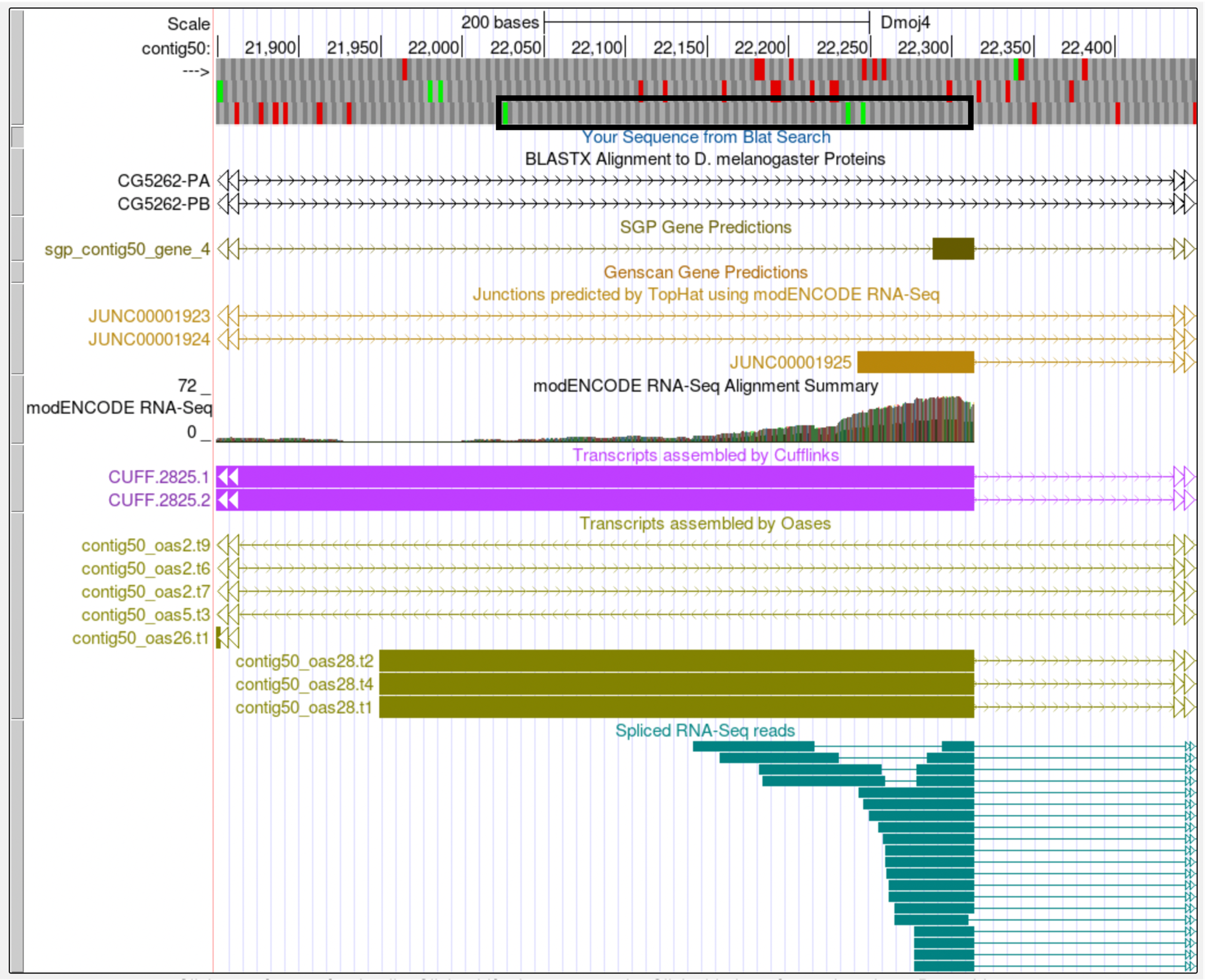
Step 6: Given the limited amount of evidence available, we will utilize any gene prediction tracks on contig50 that align well with the rest of the exons for CG31999. While the Genscan gene prediction track has two predicted exons upstream from the second exon, there is also an SGP gene prediction that is much better supported by the RNA-Seq data and TopHat splice junction predictions (Figure 24).
The Cufflinks transcripts, Oases transcripts, and spliced RNA-Seq reads all show an intron between the 5' end of the second exon and the 3' end of the predicted exon from SGP. The modENCODE RNA-Seq Alignment Summary track shows substantially more reads aligning to this region than the region that corresponds to the Genscan prediction. In addition, there is a high quality splice donor site at the end of the SGP predicted exon. While this prediction is not for an initial exon, the evidence is stronger here than the location suggested by the Genscan prediction. Therefore, we will use the +3 open reading frame from this SGP gene prediction to create the first coding exon for CG31999 (Figure 25).
The coordinates for the initial exon of CG31999 will be 22,026-22,314. Use of frame +3 creates a matching phase 1 splice donor (22,315-22,316). While the length of this initial exon is much larger than the corresponding exon from D. melanogaster (96 vs. 24 amino acids), we have exhausted all other possibilities for finding this CDS based on sequence or length conservation. Based on the available computational evidence shown in the Genome Browser, this is our best guess as to where the initial exon of CG31999 is located in D. mojavensis. Until additional evidence becomes available, we will not be able to prove or disprove the existence of this exon in D. mojavensis.
|
|
If RNA-Seq data is unavailable or if it does not support a computational gene prediction, the next best option is to use the gene prediction closest to the adjacent annotated exon (after you have verified that the splice sites for the predicted exon have the correct phase). If there are no open reading frames that have the correct phase or lack the start codon (when you are looking for an initial exon) or stop codon (when you are looking for a terminal exon), then the isoform might not exist in the species that you are trying to annotate. However, before making this decision, you should attempt all the annotation techniques described above and consult with Wilson or Chris. |
Exons of small length
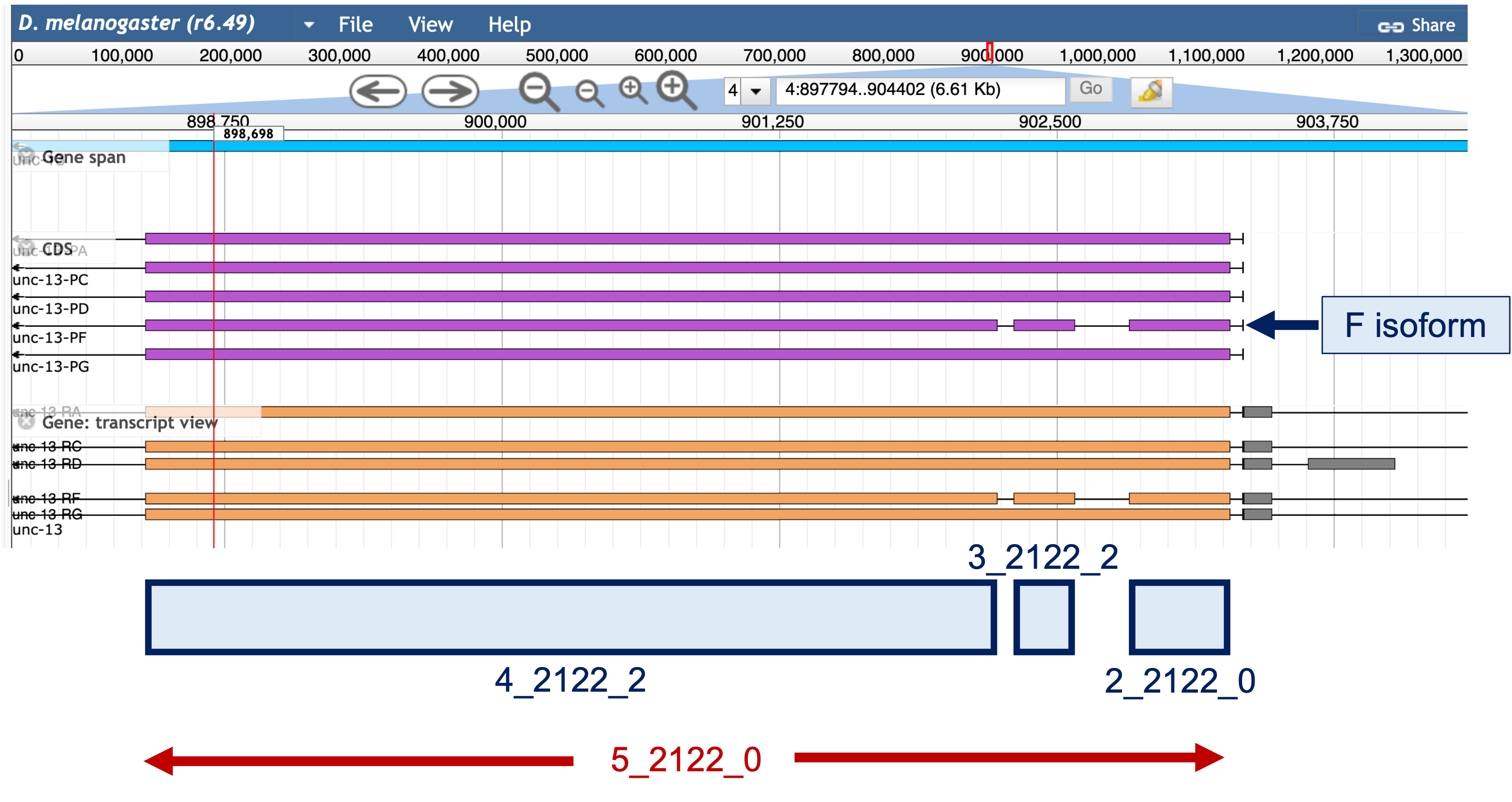
Small exons might be conserved, but due to their size, are difficult to locate with just a simple BLAST analysis. We will use CDS 1_2119_0, the initial exon for isoforms A, C, D, F, and G of unc-13, to demonstrate the step-by-step strategy for annotating small exons below.
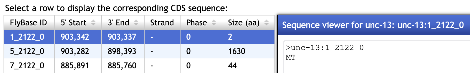
Step 1: The Gene Record Finder entry for unc-13 shows that CDS 1_2119_0 only consists of 2 amino acids (Figure 26). We will try to find this CDS on contig12 of D. mojavensis.
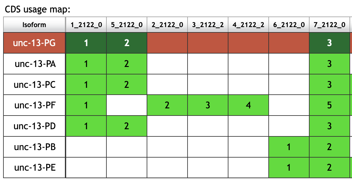
The CDS usage map shows that there is a single CDS (5_2119_0) between CDS 1_2119_0 and 7_2119_0 in the A, C, D, and G isoforms. By contrast, there are three CDS’s (2_2119_0, 3_2119_2, and 4_2119_2) between CDS 1_2119_0 and 7_2119_0 in the F isoform (Figure 27). However, closer examination of CDS coordinates and the JBrowse view shows that these three CDS’s in the F isoform actually overlaps completely with the much larger CDS in the A, C, D, and G isoforms (Figure 28).
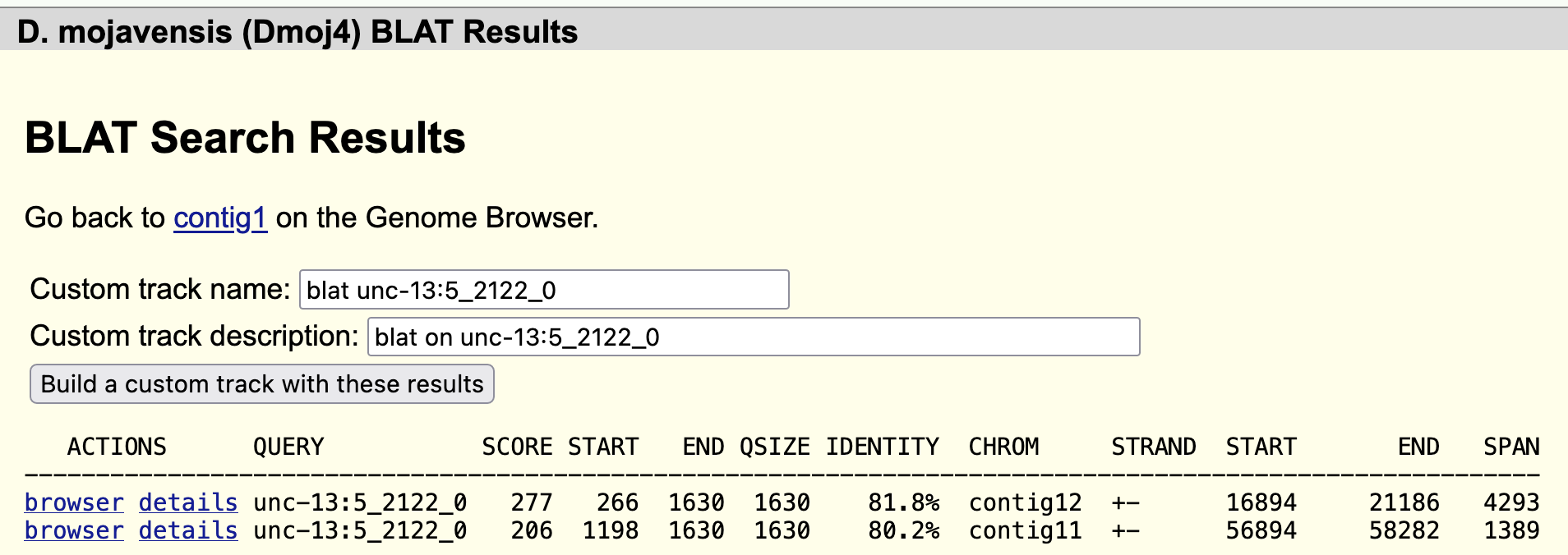
To narrow down the search region, we will use BLAT to locate CDS 5_2119_0 (i.e., the second CDS of the A, C, D, and G isoforms). BLAT found matches in both contigs 11 and 12. Because the match to contig11 is at the end of the contig (Figure 29), we will focus our efforts searching for the first CDS in contig12.
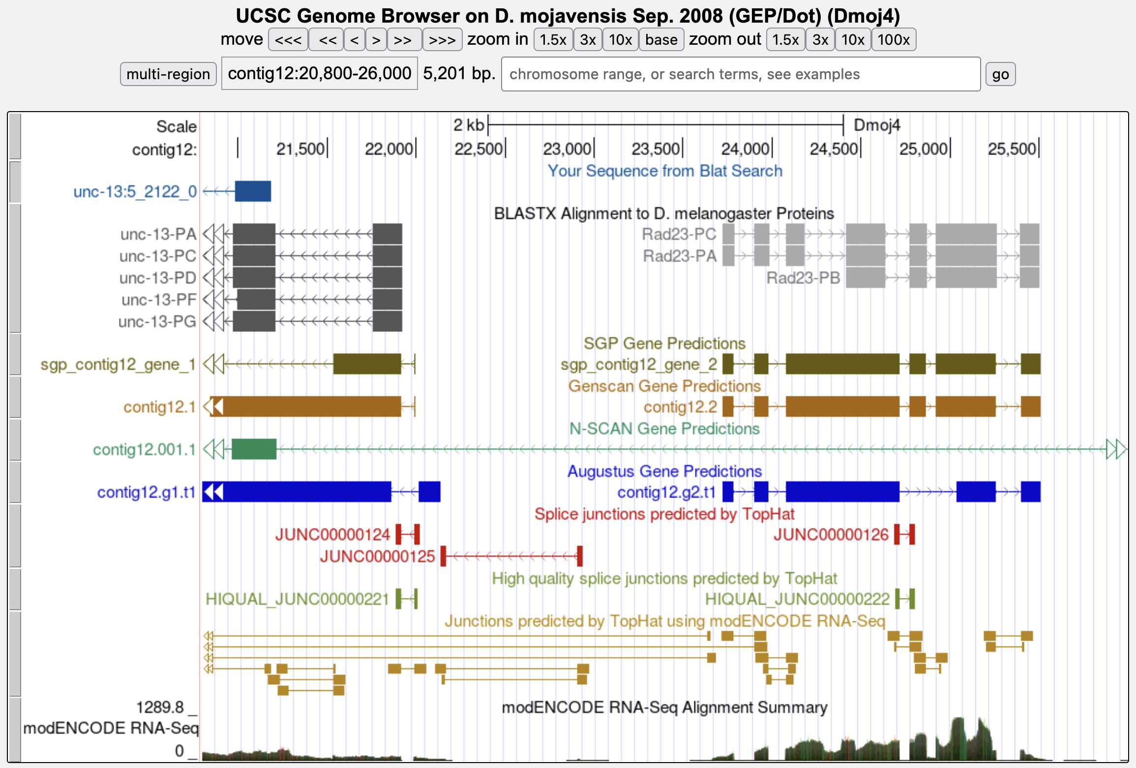
Step 2: Examination of the blastx track for contig12 shows that Rad23 is located next to unc-13 (Figure 30). Because nested genes are rare in Drosophila, we can define one end of the search boundary based on the start of the first CDS of Rad23. The tblastn alignment of the first CDS of Rad23 (1_2299_0) against contig12 shows that the CDS begins at 23,720.
The results in Figure 29 shows that the first 265 residues of CDS 5_2119_0 are missing from the BLAT alignment to contig12. Extrapolating the size of the missing region from the beginning of the alignment, we expect to find the beginning of CDS 5_2119_0 at around 21,981 (i.e., 21186 + (265 × 3)). A closer examination of this region indicates that there is a splice acceptor site at 21,917-21,916 that is strongly supported by the Genscan and SGP gene predictions as well as the TopHat junctions and RNA-Seq data (Figure 31).
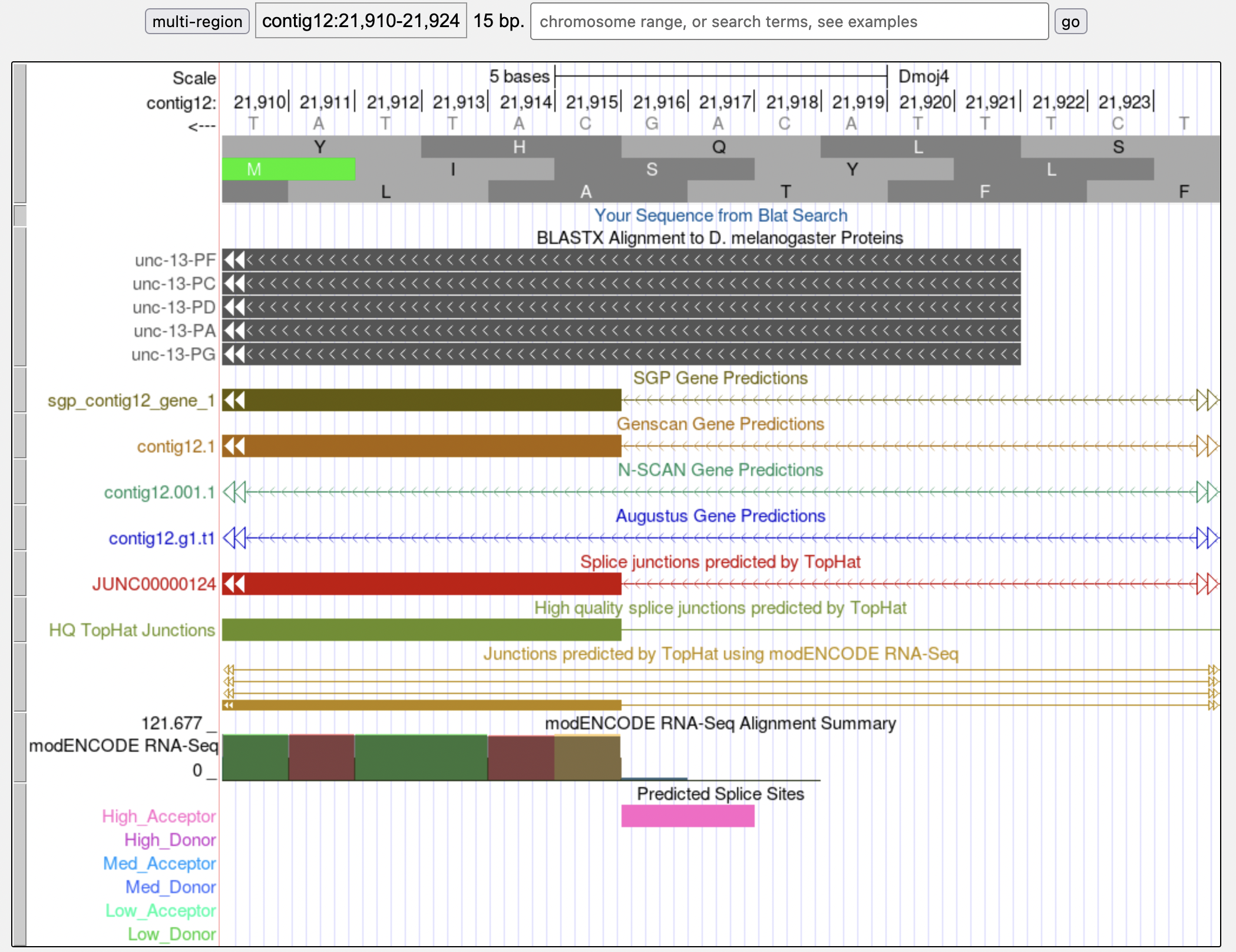
Based on the results of the tblastn search of CDS 5_2119_0 against contig12, we know that the CDS is in frame -1, which means that the splice acceptor site at 21,917-21,916 is in phase 0. This also means that the phase of the donor site for the first CDS of unc-13 (CDS 1_2119_0) is in phase 0.
Collectively, our analysis helps narrow down the first CDS to the region 21,916-23,719. In addition, this initial CDS must begin with a methionine and ends with a phase 0 donor site.
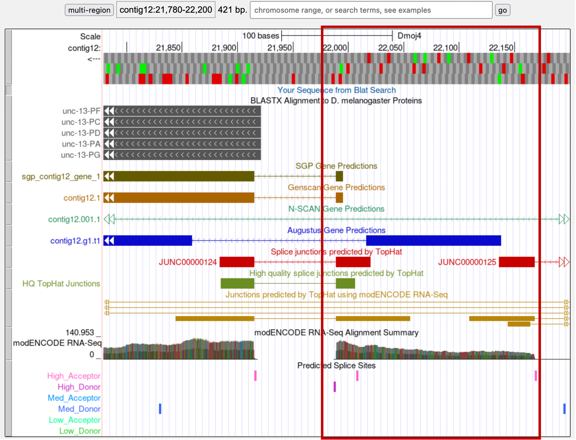
Step 3: Looking at this region in the Genome Browser, we can see that the region around 21,990-22,168 have strong evidence from RNA-Seq expression data and computational predictions: a few gene and splice junction predictors have demarcated the same CDS boundaries, and the RNA-Seq data fits these boundaries as well (Figure 32).
Zooming into this region, we see that the amino acid sequence is MT (in frame -2), the same as the D. melanogaster ortholog. This coding exon has a phase 0 splice donor, which is compatible with the phase 0 acceptor we have previously identified (Figure 33).
According to the RNA-Seq read coverage, there are regions upstream of this CDS that is part of this gene. However, given the presence of in-frame stop codons (and the gene model in D. melanogaster), these upstream regions are likely part of the 5' UTR’s of unc-13.
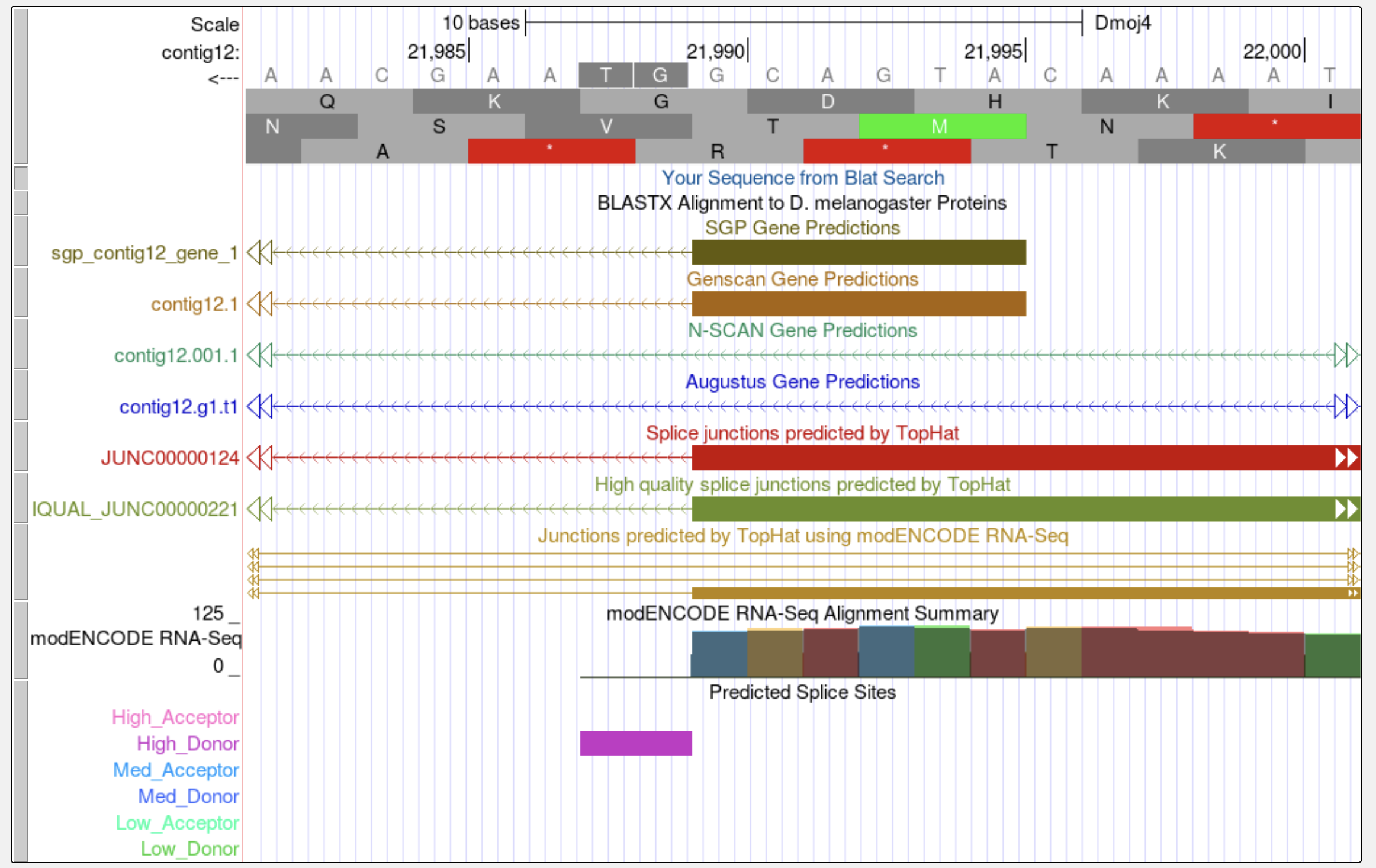
Alternate approach: Using the search boundary we have previously defined, we can also search for candidate open reading frames using the Small Exons Finder. With our search criteria, as outlined in Figure 34, the Small Exons Finder identified three candidates. Only one of these candidates (at 21,990-21,995) contains the same amino acid sequence as the orthologous CDS in D. melanogaster (MT). When we investigate the three possible candidates using the Genome Browser, we find that the only feature that is supported by multiple gene predictors and RNA-Seq data spans from 21,990 to 21,995. Consequently, the best candidate for the orthologous unc-13 CDS 1_2119_0 is located at 21,990-21,995.